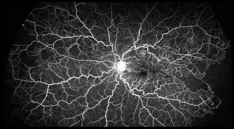
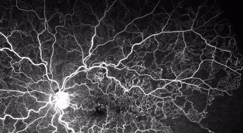
The the image shown above is of a 68-year-old male with superior hemi-retinal vein occlusion and was taken with our Optos ultra-widefield (UWF) Angiography System. Note all of the collaterals throughout the superior retina. Visual Acuity is 20/150. Unfortunately, the right eye is Light Perception from an Ischemic CRVO.
In the past, a photo like this was almost impossible. Imaging the entire retina required moving the camera from this angle to that angle in order to obtain five to seven mapping photos that we would merge together in order to obtain the best possible image of the eye as a whole. This could be challenging and frustrating for both the doctor and the patient. Since adding the Optos California and its UWF imaging capabilities, we have been able to easily increase our views of the retina and decrease patient imaging time. Speaking of the imaging device, Dr. Dale Brown said, "The fluorescein angiography capability of the Optos allows me to see more peripheral retinal nonperfusion and neovascularization than the standard method of angiography." With its use of autofluorescence, ICG and fluorescein angiography, the photos of the eye the Optos California produces are both stunning and revolutionary. The benefits for us, the doctors, are many. Dr. Jeffrey Fuller commented that, "The ability to evaluate the peripheral vasculature has been very helpful in identifying subtle neovascularization and ischemia. This has been useful in patients with PDR, BRVO, and Sickle Cell Retinopathy especially. The camera is very helpful in guiding the placement of the thermal laser into those highly damaged areas of ischemia."
0 Comments
Leave a Reply. |
AuthorThe staff and doctors at VisionAmerica are committed to providing relevant information for you, your patients and your practice. We hope you find the information in our blog post helpful. Archives
August 2019
Categories |
 RSS Feed
