|
7/18/2017 0 Comments Photo of the Month - CMEThe photos below are of a recent case treated at VisionAmerica. The diagnosis was cystoid macular edema (CME), but, as you can see, this was not your run of the mill CME. 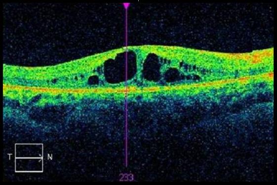  Quick Synopsis
Diagnosis: CME associated with Retinitis Pigmentosa Discussion: The OCT and Fundus photo are from the same patient. CME associated with Retinitis Pigmentosa, has been widely reported. One study reported a prevalence of almost 50%. With the loss of peripheral vision associated with RP, any reduction in central acuity is obviously devastating. Treatment: Carbonic Anhydrase Inhibitors (topical or systemic) have been show to provide an objective improvement in foveal thickness.
0 Comments
Leave a Reply. |
AuthorThe staff and doctors at VisionAmerica are committed to providing relevant information for you, your patients and your practice. We hope you find the information in our blog post helpful. Archives
August 2019
Categories |
 RSS Feed
