|
4/16/2019 0 Comments April Photo of the Month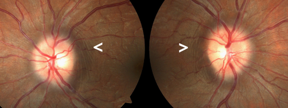 The image above is of a recent papilledema patient with Paton's Folds.
First described by Paton and Holmes in 1911, Paton's folds are concentric retinochoroidal folds that appear around the nerve in patients with papilledema. "as the disc swells lateralwards, it displaces the retina... throwing it into a series of folds which run concentric with the edge of the disc. This lateral bulging is due to the distension of the most peripheral nerve fibers.."(1a).1 1. Paton L, Holmes G. The pathology of papilloedema: a histological study of sixty eyes. Brain. 1911;33:389-432.
0 Comments
Leave a Reply. |
AuthorThe staff and doctors at VisionAmerica are committed to providing relevant information for you, your patients and your practice. We hope you find the information in our blog post helpful. Archives
August 2019
Categories |
 RSS Feed
