|
8/22/2018 0 Comments A Follow Up on Uveal Melanoma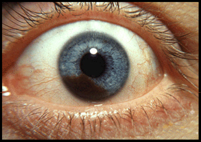 One of the highlights from our summer conference was the presentation by our own Dr. Michael Eddins on uveal melanomas. With the recent surge of interest coming out of Auburn, Alabama, we asked Dr. Eddins to provide a quick summary of his talk for those who were not able to attend. By Dr. Michael Eddins VisionAmerica Birmingham First, let me say that it was great meeting so many of you, our referring doctors, at the 2018 VisionAmerica Summer Conference! I am really looking forward to serving you and your patients! Now, onto uveal melanomas! Uveal melanomas are the most common primary malignant tumor of the eye. It has an annual incidenceof 3-10 cases per million and increases with age and peaks around age 70. Susceptibility factors include Caucasian race and light eye color. As we all know, the most common uveal melanoma is choroidal melanomas. However, there is no consistent evidence that UV exposure increases risk. With regard to location, the iris is the least common (2-4%) but carries the best prognosis. Ciliary body melanoma is the second most common (4-7%) and has the worst prognosis. In terms of the individual lesions, iris melanomas are usually small, slow-growing and rarely metastasize (3% in five years). Management typically begins with observations and serial photographs. Biopsy with fine needle aspiration can be done but does have the potential for false negatives. If growth is documented, you can consider surgical resection, plaque brachytherapy or proton beam radiation for larger lesions that are unable to be resected or even enucleation for those with diffuse spread (>50% of iris and angle) or extraocular extension. Ciliary body melanomas are more challenging. They rarely erode through the iris root or sclera and can cause cataract, subluxed lens, secondary glaucoma, uveitis, or iris neovascularization. The first sign can be dilated episcleral sentinel vessels and 50% develop metastasis despite treatment. The average survival rate with metastasis is 6 months to 1 year and treatment options include plaque brachytherapy, resection by partial lamellar sclerouvectomy, charged particle radiation, stereotactic radiosurgery, or enucleation. Choroidal Melanoma presents with the classic appearance of a pigmented dome or collar-button shaped lesion. We have all heard the To Find Small Ocular Melanomas (TFOS) mnemonic for evaluating melanomas. The signs predictive of growth include:
In terms of follow up, the typical recommendations from the Shield's group is six months after a lesion is first noted, then yearly for those with no concerning signs. For those with one-to-two signs, the recommendation is following up every4-6 months. With three or more signs, a referral to retinal oncology specialist for evaluation and possible treatment is recommended. One of the key aspects of monitoring these lesions is fundus photography and ultrasound. Color photographs are essential for evaluating for subtle growth between visits and ultrasound is critical to measure the apical height and basal diameter. As we noted in our email on July 26, 2018, the Alabama Department of Public Health has released a statement on uveal melanomas in Alabama and there are multiple efforts ongoing to build awareness. The Auburn Ocular Melanoma Facebook Page has been formed to inform those affected by the disease and build a community of support. If you have any questions about uveal melanoma, please do not hesitate to reach out to us. We are here to help!
0 Comments
Leave a Reply. |
AuthorThe staff and doctors at VisionAmerica are committed to providing relevant information for you, your patients and your practice. We hope you find the information in our blog post helpful. Archives
August 2019
Categories |
 RSS Feed
